In hospital and research facility settings, electronic analytical balance provides the critical mass measurement that is needed for delicate analyses. It is the case that reagents, samples, and medicines are weighed with the highest level of precision. Laboratory staff regard the electronic analytical balance as their helper in making the measurements, carrying out calibrations, and performing quality assurance. Besides being of great assistance in the above activities and ensuring accurate measurement in clinical diagnosis, experimental research, and drug response monitoring, electronic analytical balance also enhances overall laboratory performance and has a positive impact on the dependability of analytical results.

electronic analytical balance is commonly used in the compounding of medicinal and chemical substances in small quantities in hospital pharmacy laboratories. Mass control with high precision is critical when dealing with active pharmaceutical ingredients in micro or milligram quantities. This application helps to prepare exact doses, internal validation, and experimental drug research. By allowing for repeatable measurements, electronic analytical balance helps pharmacists and researchers in controlling formulation processes, which makes it easier to get consistency and reliability throughout hospital medication development workflows.
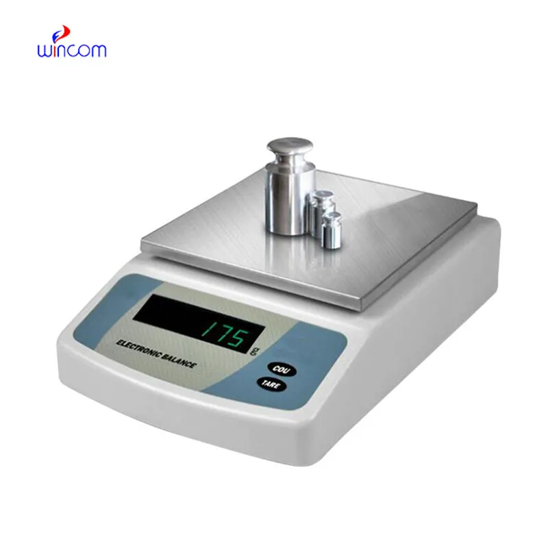
At the medical institutions that are research-driven, electronic analytical balance will change to facilitate the analytical methods with higher sensitivity that are in the pipe. The future might bring along the possibility of ultra-low mass samples being accurately measured in molecular diagnostics and sophisticated drug research. This turning development will not only enlarge the experimental capacities of hospital-based research labs but also open new fronts in medical innovation through analytics.
Most care routines for electronic analytical balance consist of planned startup and shutdown methods. Giving enough time for the warm-up process to take place guarantees that internal electronics will be operating at stable conditions. Power cuts are not allowed and this way sensitive circuits are protected. The critics of the hospitals that have implemented standard operating procedures operate daily lab analyses with a consistent measure of accuracy and at the same time prolong the life of the electronic analytical balance.
electronic analytical balance is employed in hospital labs for the reliable quality control of reagents, chemicals, and medications. Its exactness provides accurate concentrations for assays, patient treatments, and experimental protocols. The laboratory personnel regularly calibrate electronic analytical balance to rule out mistakes. Its application keeps the standard of hospital laboratories, allows the reproducibility, and builds trust in clinical and research outcomes.
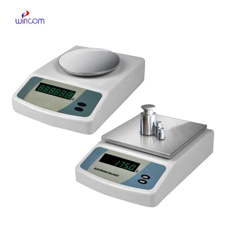
Q: What distinguishes an Analytical Balance from a precision balance? A: The analytical balances have a higher sensitivity and a finer readability for measuring masses of very small amounts. Q: Is an Analytical Balance appropriate for pharmaceutical applications? A: It is widely used for weighing active ingredient and formulation components. Q: Is it mandatory for an Analytical Balance to have a draft shield? A: Draft shields have the function to prevent air disturbances which might affect the weighing results. Q: What are the possible types of materials that can be weighed on an Analytical Balance? A: Weighing of powders, chemicals, and biological samples, as well as reference weights are the most common measurement. Q: Is it possible for several users to work with the same Analytical Balance? A: Yes, but the proper handling procedures and access controls must be strictly adhered to.
The centrifuge operates quietly and efficiently. It’s compact but surprisingly powerful, making it perfect for daily lab use.
We’ve used this centrifuge for several months now, and it has performed consistently well. The speed control and balance are excellent.
To protect the privacy of our buyers, only public service email domains like Gmail, Yahoo, and MSN will be displayed. Additionally, only a limited portion of the inquiry content will be shown.
We’re interested in your delivery bed for our maternity department. Please send detailed specifica...
We are planning to upgrade our imaging department and would like more information on your mri machin...
E-mail: [email protected]
Tel: +86-731-84176622
+86-731-84136655
Address: Rm.1507,Xinsancheng Plaza. No.58, Renmin Road(E),Changsha,Hunan,China

 af
af
 es
es
 ar
ar
 tr
tr
 sw
sw
 pt
pt
 th
th
 ur
ur
 bn
bn
 ne
ne
 vi
vi
 km
km
 lo
lo
 de
de
 ru
ru
 fi
fi
 nl
nl
 fa
fa
 fr
fr
 ko
ko