In the laboratories of hospitals and clinics, electronic benefit transfer delaware balance is responsible for the precise weighing of patient samples, reagents, and pharmaceutical ingredients. Being highly precise, it minimized sample preparation errors and that is a good support for analytical results that can be reproduced. Laboratory techs apply electronic benefit transfer delaware balance in the processes of quality control, method validation, and even daily operations. Reliable diagnostics, efficient laboratory workflows, and high-quality research and medical testing are the consequences of the accuracy and consistency maintained by electronic benefit transfer delaware balance.
electronic benefit transfer delaware balance is commonly used in the compounding of medicinal and chemical substances in small quantities in hospital pharmacy laboratories. Mass control with high precision is critical when dealing with active pharmaceutical ingredients in micro or milligram quantities. This application helps to prepare exact doses, internal validation, and experimental drug research. By allowing for repeatable measurements, electronic benefit transfer delaware balance helps pharmacists and researchers in controlling formulation processes, which makes it easier to get consistency and reliability throughout hospital medication development workflows.
electronic benefit transfer delaware balance will be an advanced generation with self-diagnostic features as the norm. Predictive monitoring of internal parts will assist laboratory teams to schedule maintenance activities in a much more efficient manner. This will lead to the continuous operation of hospital laboratories where downtime is a direct impact on both clinical workflows and research schedules.
In order to keep electronic benefit transfer delaware balance in a good condition consistent calibration practices are needed that follow hospital laboratory protocols. Scheduled calibration checks are performed to maintain the reliability of measurements during daily activities involving analysis. Conditions in the environment such as temperature and the amount of air that moves around should be kept under control so as to prevent drift. The people operating the machines should make sure that there are no sudden changes in load and that the weighing pan is not subjected to excessive force. Through adhering to controlled handling practices, electronic benefit transfer delaware balance is always trusted for pharmaceutical preparation and medical research activities.
electronic benefit transfer delaware balance plays an important role in the hospital pharmacy in the accurate formulation of medications, intravenous solutions, and compounded drugs. Even slight alterations in the weight of drugs can change the effectiveness of the drug and endanger the patient's safety. The Pharmacy Technicians rely on electronic benefit transfer delaware balance for the correct dosing and checking of the active ingredients. The tool's accuracy guarantees the dependable preparation of drugs, the observance of the rules, and the quality control of the overall hospital pharmacy activities.
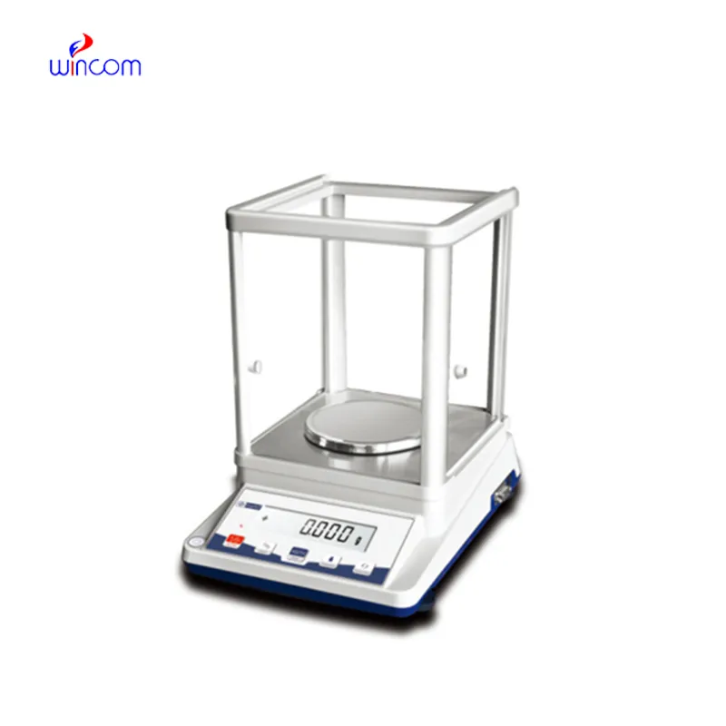
Q: What industries are the widespread users of Analytical Balances? A: Their primary application lies in laboratories, hospitals, pharmaceutical facilities, and research institutions. Q: Is it possible to measure liquids with an Analytical Balance? A: Liquids can be indirectly measured using appropriate containers. Q: What does it mean by repeatability in an Analytical Balance? A: It is the capability to provide constant results when tested under the same conditions. Q: Is it necessary for the installation of Analytical Balances to be in controlled environments? A: Controlled environments are beneficial for keeping accuracy and stability long term. Q: What is the average lifespan of an Analytical Balance? A: If taken care of and maintained properly, it will be a reliable and many years-long operating device.
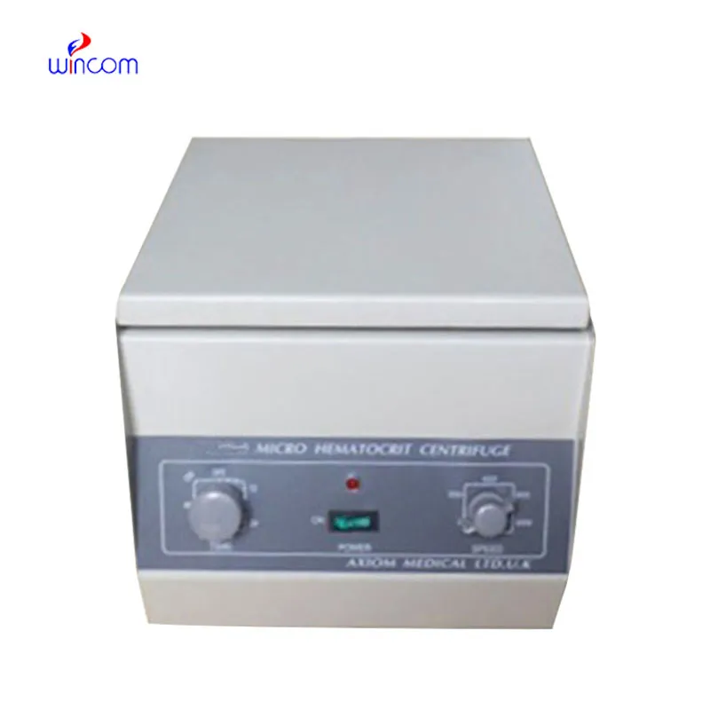
We’ve used this centrifuge for several months now, and it has performed consistently well. The speed control and balance are excellent.
The water bath performs consistently and maintains a stable temperature even during long experiments. It’s reliable and easy to operate.
To protect the privacy of our buyers, only public service email domains like Gmail, Yahoo, and MSN will be displayed. Additionally, only a limited portion of the inquiry content will be shown.
We’re looking for a reliable centrifuge for clinical testing. Can you share the technical specific...
Hello, I’m interested in your water bath for laboratory applications. Can you confirm the temperat...
E-mail: [email protected]
Tel: +86-731-84176622
+86-731-84136655
Address: Rm.1507,Xinsancheng Plaza. No.58, Renmin Road(E),Changsha,Hunan,China

 af
af
 es
es
 ar
ar
 tr
tr
 sw
sw
 pt
pt
 th
th
 ur
ur
 bn
bn
 ne
ne
 vi
vi
 km
km
 lo
lo
 de
de
 ru
ru
 fi
fi
 nl
nl
 fa
fa
 fr
fr
 ko
ko