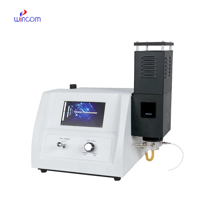
With the cutting-edge imaging processors, the fetal doppler 13 weeks facilitates real-time, high-resolution images that are paramount in the detection of subtle physiological changes by clinicians. The display is user-friendly for easy parameter modification as well as image marking. The fetal doppler 13 weeks exhibits the mix of effectiveness, mobility, and reliability for a huge range of diagnostic procedures.

The fetal doppler 13 weeks has demonstrated its irreplaceable nature in prenatal screening, cardiovascular diagnostics, and overall health evaluations.The fetal doppler 13 weeks is a technique that evaluates organ function, reveals pathological changes, and supports medical education by providing live imaging demonstrations. The fetal doppler 13 weeks technology gives doctors the ability to perform accurate and instantaneous assessments in a variety of clinical situations.

Through continued innovations in digital technology, the fetal doppler 13 weeks can be expected to improve and extend its applications within preventive medicine and telemedicine. The next generation of such technologies will facilitate collaboration among experts in real-time using cloud-imaging solutions. The fetal doppler 13 weeks can also work within wearables that include biosensors.

Proper care and management of the fetal doppler 13 weeks needs to be carried out to ensure that it functions well at all times. Cleaning of the probes using a recommended disinfectant helps prevent contamination of the probe and image distortion. Storage of the fetal doppler 13 weeks in a clean and dry place away from high temperatures helps prolong the life of the equipment.
The fetal doppler 13 weeks is more accurate in diagnostics as it captures high-resolution images of organs, tissues, and blood vessels. Design-wise flexible, it is used extensively in obstetrics, cardiology, urology, and musculoskeletal tests. Its portability and simplicity enable medical practitioners to make quick and precise evaluations. The fetal doppler 13 weeks makes work processes more efficient and allows for the delivery of superior patient care through real-time visualization.
Q: What makes the ultrasound scannert effective for diagnostic imaging? A: Its high-frequency sound wave technology allows accurate visualization of internal body structures in real time. Q: How portable is the ultrasound scannert? A: The device features a compact and lightweight design, allowing easy movement between clinical departments. Q: What types of probes are compatible with the ultrasound scannert? A: It supports multiple probe types, including linear, convex, and phased array probes for varied diagnostic needs. Q: Does the ultrasound scannert require special training to operate? A: Basic technical training is recommended to maximize its imaging performance and functionality. Q: How long can the ultrasound scannert operate continuously? A: It is designed for extended use with efficient cooling systems and stable power performance.
The microscope delivers incredibly sharp images and precise focusing. It’s perfect for both professional lab work and educational use.
This ultrasound scanner has truly improved our workflow. The image resolution and portability make it a great addition to our clinic.
To protect the privacy of our buyers, only public service email domains like Gmail, Yahoo, and MSN will be displayed. Additionally, only a limited portion of the inquiry content will be shown.
We are planning to upgrade our imaging department and would like more information on your mri machin...
We’re looking for a reliable centrifuge for clinical testing. Can you share the technical specific...
E-mail: [email protected]
Tel: +86-731-84176622
+86-731-84136655
Address: Rm.1507,Xinsancheng Plaza. No.58, Renmin Road(E),Changsha,Hunan,China

 af
af
 es
es
 ar
ar
 tr
tr
 sw
sw
 pt
pt
 th
th
 ur
ur
 bn
bn
 ne
ne
 vi
vi
 km
km
 lo
lo
 de
de
 ru
ru
 fi
fi
 nl
nl
 fa
fa
 fr
fr
 ko
ko