The fetal doppler heartbeat works by integrating cutting-edge image optimization features that reduce artifacts and improve detail recognition thereby increasing diagnostic accuracy. It caters to multilingual users and sets for personal usability requirements for global needs. The device provides imaging of the same quality regardless of the patient type or clinical condition.

The fetal doppler heartbeat has a very wide use in radiology where it supports the guidance of non-invasive procedures. It is very important in gynecology where it is allowed to conduct reproductive system evaluations. In orthopedics, the fetal doppler heartbeat helps visualize muscles, joints, and tendons ensuring correct diagnostic interpretation. It is this very nature of versatility that makes it a must-have for imaging during procedures in real time.

Through continued innovations in digital technology, the fetal doppler heartbeat can be expected to improve and extend its applications within preventive medicine and telemedicine. The next generation of such technologies will facilitate collaboration among experts in real-time using cloud-imaging solutions. The fetal doppler heartbeat can also work within wearables that include biosensors.

The daily upkeep of the fetal doppler heartbeat involves cleaning, inspection, and proper storage. The removal of the gel residue from the probes should be accomplished as soon as the analysis has been carried out. The cooling vents of the device should always be unblocked. The fetal doppler heartbeat needs annual professional servicing in order to remain accurate.
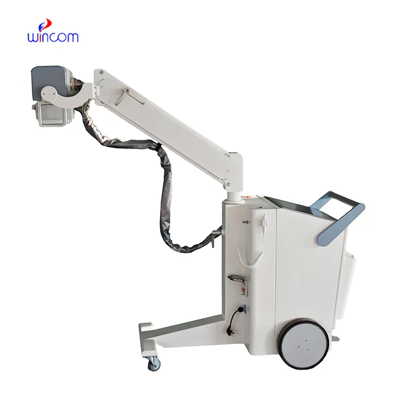
The fetal doppler heartbeat is an essential diagnostic modality in the modern healthcare system, permitting the non-invasive imaging of internal organs and tissues. By transmitting sound waves and reading their echoes, it provides real-time data on physiology. The fetal doppler heartbeat makes precise diagnoses feasible in all specialties, improving clinical decision-making and patient confidence.
Q: What makes the ultrasound scannert effective for diagnostic imaging? A: Its high-frequency sound wave technology allows accurate visualization of internal body structures in real time. Q: How portable is the ultrasound scannert? A: The device features a compact and lightweight design, allowing easy movement between clinical departments. Q: What types of probes are compatible with the ultrasound scannert? A: It supports multiple probe types, including linear, convex, and phased array probes for varied diagnostic needs. Q: Does the ultrasound scannert require special training to operate? A: Basic technical training is recommended to maximize its imaging performance and functionality. Q: How long can the ultrasound scannert operate continuously? A: It is designed for extended use with efficient cooling systems and stable power performance.
We’ve used this centrifuge for several months now, and it has performed consistently well. The speed control and balance are excellent.
The delivery bed is well-designed and reliable. Our staff finds it simple to operate, and patients feel comfortable using it.
To protect the privacy of our buyers, only public service email domains like Gmail, Yahoo, and MSN will be displayed. Additionally, only a limited portion of the inquiry content will be shown.
We’re interested in your delivery bed for our maternity department. Please send detailed specifica...
Hello, I’m interested in your water bath for laboratory applications. Can you confirm the temperat...
E-mail: [email protected]
Tel: +86-731-84176622
+86-731-84136655
Address: Rm.1507,Xinsancheng Plaza. No.58, Renmin Road(E),Changsha,Hunan,China

 af
af
 es
es
 ar
ar
 tr
tr
 sw
sw
 pt
pt
 th
th
 ur
ur
 bn
bn
 ne
ne
 vi
vi
 km
km
 lo
lo
 de
de
 ru
ru
 fi
fi
 nl
nl
 fa
fa
 fr
fr
 ko
ko