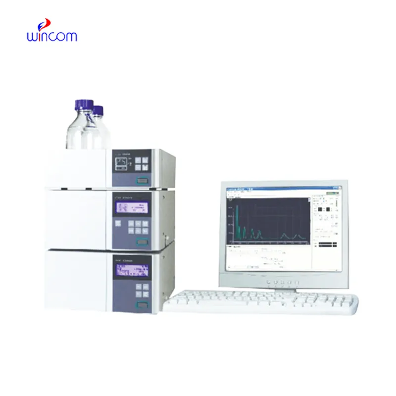
hplc analyse offers high resolution separation of complex samples in clinical, pharmaceutical, and hospital laboratories, thereby supporting advanced laboratory workflows. It allows performing an in-depth analysis of drugs, metabolites, and small biomolecules. hplc analyse is used by laboratory staff for research validation, patient monitoring, and method development. Its precision, speed, and adaptability make analytical efficiency greater and at the same time, make consistent and reproducible results which in turn, strengthen laboratory operations in the areas of healthcare and scientific environments.
hplc analyse finds use in clinical toxicology laboratories to pinpoint and measure the amounts of possible poisons or drugs in abuse samples taken from patients. It is based on the separation of the various substances from complex mixtures like blood or urine, and that information is very important for the hospital doctors, who will then diagnose the case, decide on the treatment and monitor the patient’s safety.
The hplc analyse scenario predicted for hospital labs is all about the automated sample handling systems and the digital data analysis. Cutting-edge detectors along with AI-based interpretation are going to double the accuracy and the amount of the processed samples. All this will lead to major hospitals using hplc analyse more and more for fast testing of patients, monitoring of treatments, and, with the help of research, unlocking the potential of their individual patients thus making medicine less and lab work more efficient.
The effectiveness of a laboratory is determined by the proper maintenance of hplc analyse. If the pump seals are regularly cleaned, the flow rates are monitored, and the usage of incompatible solvents is avoided then damage to the laboratory equipment can be prevented. It is essential for the technicians to carefully examine the columns, detectors, and tubing and in case of any sign of wear to conduct the scheduled calibration. Keeping hplc analyse in their best condition guarantees reproducibility, lowers the risk of equipment breakdown, and provides continuous performance for both hospital tests and experiments.
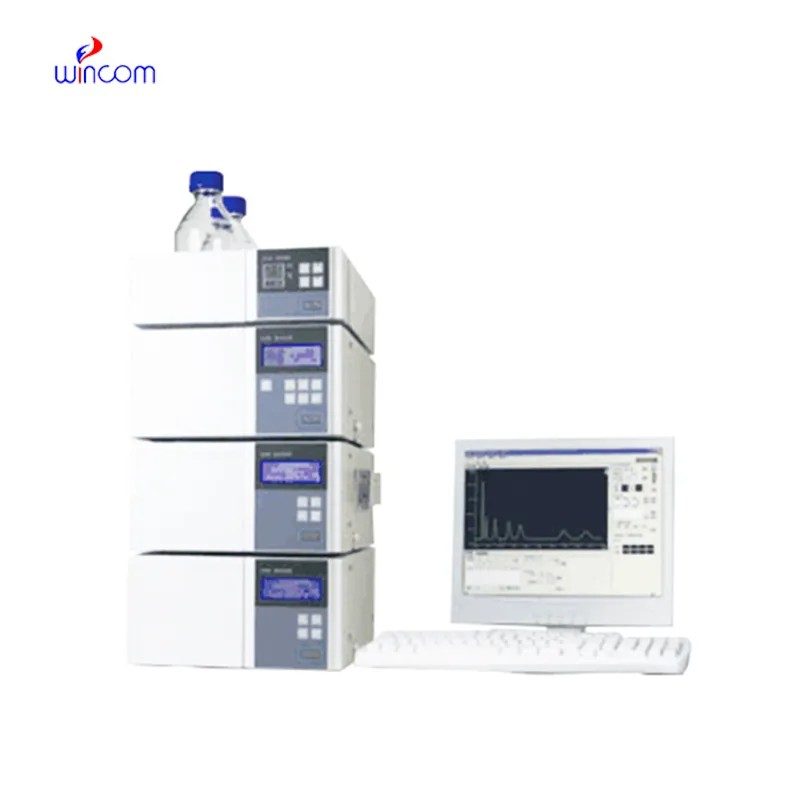
hplc analyse is commonly employed in laboratories to separate, identify, and quantify chemical compounds. The sample mixture is put through the columns along with the stationary phases and the different components interact with the stationary phase, thus the separation is done accurately. This process not only gives high resolution but also reproducibility thus it is a must-have tool for the research works in the area of drugs, pollution, and food control. Subsequently, when coupled with sensitive detectors, hplc analyse facilitates the precise measurement of minor concentrations. The method versatility produces so much that it has become a necessity in a routine analysis and complex research applications where it is positioned as an essential instrument in contemporary analytical chemistry and experimental workflows.
Q: Do you need special training for HPLC operation? A: The answer is yes, training is a prerequisite to accurately and safely using pumps, columns, and detectors. Q: What type of maintenance does HPLC have? A: It requires cleaning, flushing, and inspection of all components as well as calibrating. Q: Is it possible to use HPLC in drug monitoring? A: Sure, it is a common practice in hospitals to monitor the levels of therapeutic drugs and also to identify metabolites in the samples taken from the patients. Q: What is the duration of analysis using HPLC in a typical case? A: The analysis time can range from a few minutes to more than an hour depending on the nature of the sample and the kind of column used. Q: Is HPLC a good choice for environmental testing? A: Yes, it can be used to find out the presence of pollutants, pesticides, and other harmful substances in water, soil, and air samples.
I’ve used several microscopes before, but this one stands out for its sturdy design and smooth magnification control.
This x-ray machine is reliable and easy to operate. Our technicians appreciate how quickly it processes scans, saving valuable time during busy patient hours.
To protect the privacy of our buyers, only public service email domains like Gmail, Yahoo, and MSN will be displayed. Additionally, only a limited portion of the inquiry content will be shown.
Hello, I’m interested in your water bath for laboratory applications. Can you confirm the temperat...
We’re looking for a reliable centrifuge for clinical testing. Can you share the technical specific...
E-mail: [email protected]
Tel: +86-731-84176622
+86-731-84136655
Address: Rm.1507,Xinsancheng Plaza. No.58, Renmin Road(E),Changsha,Hunan,China

 af
af
 es
es
 ar
ar
 tr
tr
 sw
sw
 pt
pt
 th
th
 ur
ur
 bn
bn
 ne
ne
 vi
vi
 km
km
 lo
lo
 de
de
 ru
ru
 fi
fi
 nl
nl
 fa
fa
 fr
fr
 ko
ko