hplc-ms is a critical technique to obtain analytical information in studies of medicines, clinical samples, and biochemistry. It isolates compounds according to their chemical characteristics, generating reproducible analytical results. Laboratory scientists use hplc-ms to perform drug stability tests, monitor patient biomarkers, and find impurities. Its very high accuracy and flexibility allow thorough sample analysis in research, hospital, and clinical laboratory environments, thus becoming a fundamental device for assuring precision in both experimental and diagnostic results.
The hospital laboratory technicians employ hplc-ms to quantify the quantity of proteins or peptides. This assists in the research of biomarkers, immunotherapy studies, and analysis of responses induced by novel therapies among patients. Its accuracy and sensitivity enable the obtaining of correct results, hence aiding superior research.
The instruments for hplc-ms of the future will be equipped with separation methods in multiple dimensions and fully automated sample preparation. The detection of trace amounts of metabolites, drugs, and biomarkers will be so accurate that hospitals and clinical laboratories will be the first to reap the benefits. The applications of hplc-ms in the future will greatly help in complex diagnostics, research studies, and laboratory efficiency.
Systematic cleaning, pressure monitoring, and timely worn parts replacement are among the measures to be taken in the hospital laboratories to keep hplc-ms under control. Laboratory staff must ensure the observance of the suggested operating conditions, avoid the formation of air bubbles in the system, and check for proper solvent compatibility. Regular maintenance maintains the performance of the column, avoids contamination, and allows the analysis to be precise and reproducible, thereby benefiting not only routine patient testing but also experimental research.
hplc-ms is employed by laboratories in hospitals and research centers to keep control over their analytical quality in a manner that is non-stop. It works by separations of different chemicals in complex mixtures, pinpointing the impurities, and very accurately quantifying the concentrations. Technicians in the laboratory depend on hplc-ms for the purposes of method verification, calibration, and validation of techniques for analysis. It is in clinical and pharmaceutical labs that the instrument changes the generated data into accurate and reproducible forms. Its high-resolution separation capacity is utilized by both modern testing and up-to-date research projects. hplc-ms is given the credit of being the backbone instrument in laboratory operations by providing detailed results that are consistent, thus being the source of reliable analysis and supporting the whole medical and experimental research by maintaining its integrity.
Q: What is the sample preparation for HPLC? A: For the most part, samples should be filtered, diluted, or subjected to solvent extraction in order to avoid column clogs and have the results be accurate Q: Is HPLC able to pick trace-level compounds? A: With the right detectors, it can pick up such substances in extremely small amounts with high sensitivity. Q: Is HPLC a method that can be applied to analysis of proteins? A: Yes, particularly if one employs size-exclusion and reversed-phase columns for protein, peptide, and biomolecule separation. Q: What is the process of calibrating HPLC? A: The process is done by taking standards of known concentrations that are the same as the one in the sample and using them to check the performance of the column and the accuracy of the detector. Q: Are particular solvents needed for HPLC? A: Yes, the solvents used need to be compatible with the type of the column and the detectors to prevent any damage or interference in the analysis process.
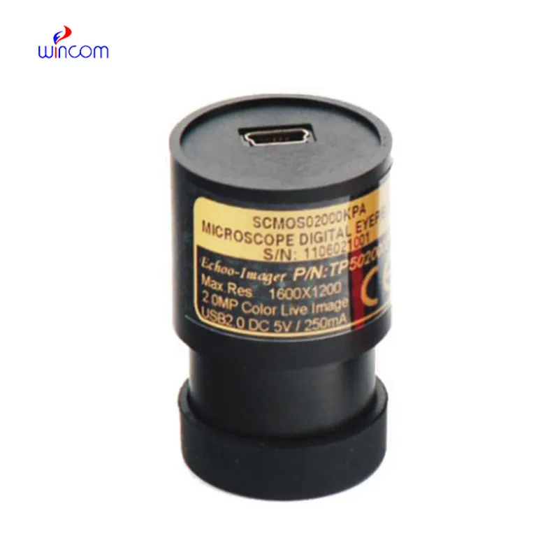
I’ve used several microscopes before, but this one stands out for its sturdy design and smooth magnification control.
The microscope delivers incredibly sharp images and precise focusing. It’s perfect for both professional lab work and educational use.
To protect the privacy of our buyers, only public service email domains like Gmail, Yahoo, and MSN will be displayed. Additionally, only a limited portion of the inquiry content will be shown.
We’re interested in your delivery bed for our maternity department. Please send detailed specifica...
We’re currently sourcing an ultrasound scanner for hospital use. Please send product specification...
E-mail: [email protected]
Tel: +86-731-84176622
+86-731-84136655
Address: Rm.1507,Xinsancheng Plaza. No.58, Renmin Road(E),Changsha,Hunan,China

 af
af
 es
es
 ar
ar
 tr
tr
 sw
sw
 pt
pt
 th
th
 ur
ur
 bn
bn
 ne
ne
 vi
vi
 km
km
 lo
lo
 de
de
 ru
ru
 fi
fi
 nl
nl
 fa
fa
 fr
fr
 ko
ko