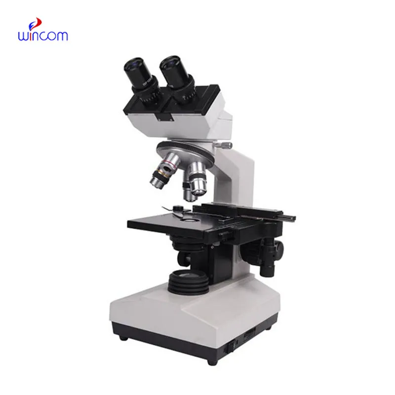
The microscope for algae observation in environmental studies pairs simple controls and precise optical alignment for smooth switching between magnifications. distortion is minimized and depth perception is enhanced with quality glass lenses. a sturdy frame and vibration-dampening base contribute to stable imaging, even under extensive use. The microscope for algae observation in environmental studies features integral LED illumination with adjustable intensity for precise light control, providing crisp and clear viewing of biological or material samples for research and educational studies.
The microscope for algae observation in environmental studies is today a vital component of sectors that involve precise observation. In schools, it promotes scientific investigation by allowing students to observe living cells and microscopic organisms. Biomedical professionals apply the microscope for algae observation in environmental studies in diagnostic testing of tissues and histological analysis. The microscope for algae observation in environmental studies is also applied in industrial testing, where engineers study welds, coatings, and fractures. The microscope for algae observation in environmental studies assists environmental scientists in studying microorganisms in soil and water environments, ensuring sustainability and preservation.

With the progress of technology, the microscope for algae observation in environmental studies will turn into a smarter and more interactive research tool. Compatibility with AI will allow it to detect patterns, recognize anomalies, and measure data automatically. The microscope for algae observation in environmental studies will also make remote diagnostics possible, where the samples from every corner of the world can be diagnosed remotely by specialists. Advances in imaging sensors and optical systems will provide better depth resolution and faster capture rates. These will expand the uses of the microscope for algae observation in environmental studies in medicine, nanotechnology, and education.

Maintenance of the microscope for algae observation in environmental studies involves regular cleaning and preventive inspection. Always start by making sure all lenses and eyepieces are clean of dust before observing. Avoid subjecting the microscope for algae observation in environmental studies to extreme temperatures or humidity levels. Clean immersion lenses after each session and remove all the slides from the stage. Keep the microscope for algae observation in environmental studies covered when not in use to protect it from contaminants. Engage professional maintenance every year to inspect optical alignment and ensure there is smooth mechanical running.
The microscope for algae observation in environmental studies is a cornerstone of scientific discovery, allowing exact observation of objects too small for the human eye. From freshman biology to medical diagnostics and materials science, the microscope for algae observation in environmental studies allows samples to be observed extensively at any level of magnification. It uses sophisticated optics and illumination to produce sharp, defining images. More recent models involve cameras and computer software to decode data in real time, allowing scientists to gather and share microscopic observations more rapidly and accurately.
Q: How do environmental conditions affect a microscope? A: Excessive heat, moisture, or dust can damage optical and mechanical components, so the microscope should be used in a clean, controlled environment. Q: Can a microscope capture images or videos? A: Many modern microscope models include digital cameras that enable high-resolution image and video capture for documentation or analysis. Q: What training is required to operate a microscope? A: Basic understanding of optics and focusing principles is recommended, though most educational microscopes are designed for simple, intuitive use. Q: Why is regular maintenance important for a microscope? A: Regular maintenance prevents dust buildup, mechanical wear, and misalignment, ensuring consistent performance and image clarity. Q: Can a microscope be used outside the laboratory? A: Portable and handheld microscope models are available for field studies, allowing researchers to observe and analyze samples on site.
The hospital bed is well-designed and very practical. Patients find it comfortable, and nurses appreciate how simple it is to operate.
This x-ray machine is reliable and easy to operate. Our technicians appreciate how quickly it processes scans, saving valuable time during busy patient hours.
To protect the privacy of our buyers, only public service email domains like Gmail, Yahoo, and MSN will be displayed. Additionally, only a limited portion of the inquiry content will be shown.
We’re looking for a reliable centrifuge for clinical testing. Can you share the technical specific...
We’re interested in your delivery bed for our maternity department. Please send detailed specifica...
E-mail: [email protected]
Tel: +86-731-84176622
+86-731-84136655
Address: Rm.1507,Xinsancheng Plaza. No.58, Renmin Road(E),Changsha,Hunan,China

 af
af
 es
es
 ar
ar
 tr
tr
 sw
sw
 pt
pt
 th
th
 ur
ur
 bn
bn
 ne
ne
 vi
vi
 km
km
 lo
lo
 de
de
 ru
ru
 fi
fi
 nl
nl
 fa
fa
 fr
fr
 ko
ko