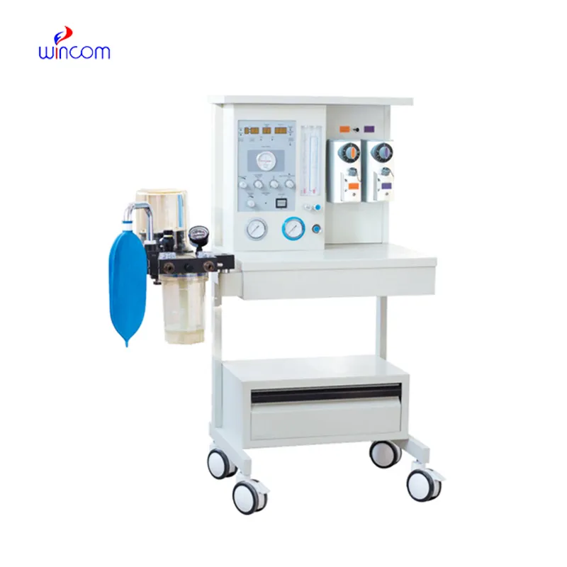
mri compatible anesthesia machine is capable of perfecting the anesthetic gas mixtures and at the same time providing mechanical ventilation for the patients during the surgical process. Its monitoring systems keep a check on the various vital parameters like oxygen saturation, airway pressure, and tidal volume. This device is widely in use in hospitals and clinical laboratories for the exact purpose of keeping sedation and respiratory function under control. The collaboration of alarm systems, flow controls that can be modified, and a real-time display permits anesthesiologists to react without delay to physiological changes. mri compatible anesthesia machine offers safe and effective anesthesia delivery which is why it is considered a must-have in operating rooms, emergency care units, and research places.
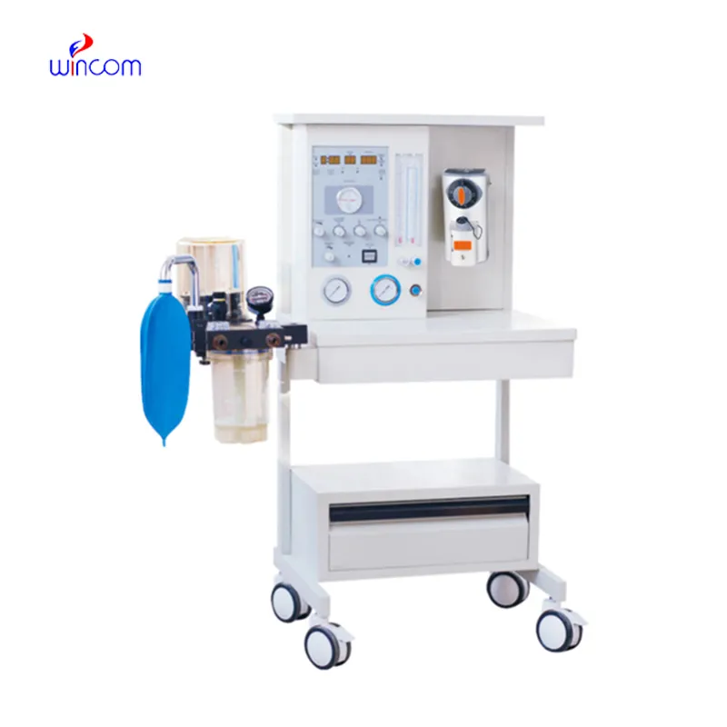
In the pediatric wards, mri compatible anesthesia machine is the method of choice for the provision of anesthesia according to children's physiological characteristics. The pediatric population is often subjected to meticulous ventilation and anesthetic dosage control because of their small airways and high sensitivity. The machine contributes to the maintenance of gentle ventilation and precise gas delivery during operations. Its monitoring feature is a constant companion to the medical practitioners who are checking the respiration rates and the oxygen concentrations. This use case points out the necessity of mri compatible anesthesia machine in guaranteeing the safety of young patients when they get anesthetized in hospitals.
Sustainability is becoming more and more an important factor to be considered in the future of mri compatible anesthesia machine. Manufacturers might concentrate on waste gas anesthetics elimination and energy efficiency enhancements. hospitals are getting more and more conscious of environmental effects and operating expenses. Future apparatus may couple state-of-the-art gas recycling systems and low-flow anesthesia capabilities. These alterations could contribute to the responsible resources usage and at the same time, keep up with clinical performance. The progress of mri compatible anesthesia machine is expected to be in sync with the larger hospital sustainability projects.
The gas delivery parts of mri compatible anesthesia machine maintenance need to be very careful. It is a must to have the oxygen sensors and gas flow regulators tested frequently to verify their correctness. Performance issues can be caused if the hose or connector leaks are not detected and fixed soon. In the operating theaters, dependable gas supply is a means of securing the patients' safety during the surgeries. Periodic testing makes sure that the apparatus is always operating properly under different clinical scenarios.
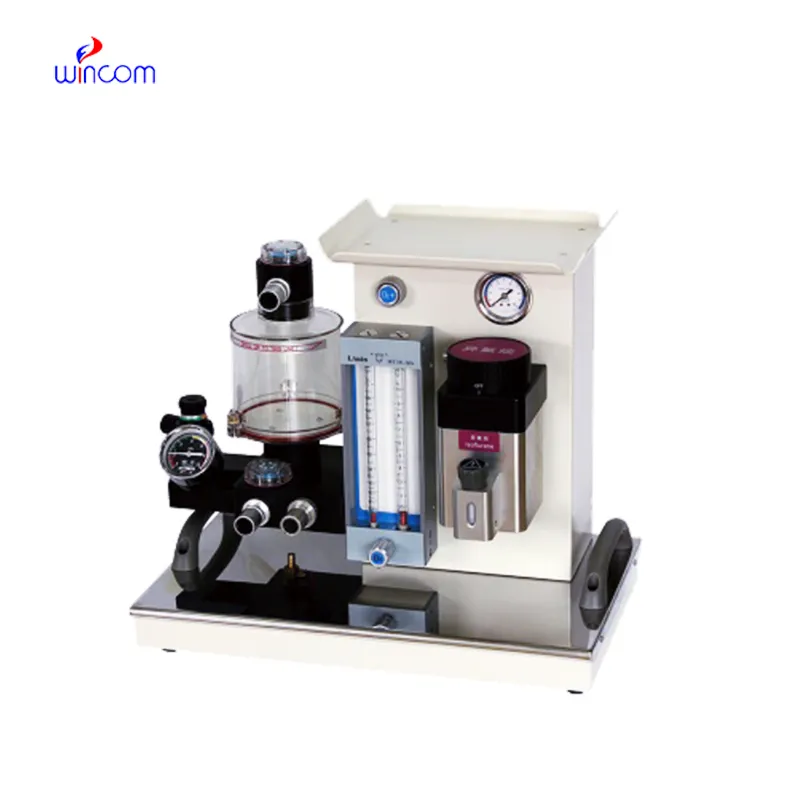
mri compatible anesthesia machine is a crucial medical apparatus used in hospitals, intended to administer accurate anesthetic gases and at the same time, allow patient ventilation during surgery. It incorporates monitoring systems which constantly measure the oxygen levels, respiratory rate, and airway pressures, thus ensuring patient safety during the entire procedure. The device, which is made up of vaporizers, flow meters, and alarms, allows anesthesiologists to control sedation uniformly and react to different physiological situations. Its use goes from operating rooms to emergency departments and laboratory-based clinical studies, thus providing consistent performance for efficient anesthesia administration and facilitating good patient management in various hospital environments.
Q: What is the main function of an anesthesia machine? A: Controlled anesthetic gases are delivered and the patient is ventilated during medical procedures. Q: Where is the anesthesia machine frequently used? A: Hospitals mainly use it in their operating rooms, emergency units, and intensive care departments. Q: Who operates the anesthesia machine during surgery? A: Trained anesthesiologists or anesthesia technicians are responsible for operating the device. Q: Can the anesthesia machine assist the breathing of a patient? A: Yes, by providing mechanical ventilation support when spontaneous breathing is insufficient. Q: Does the device monitor respiratory parameters? A: It monitors airflow, pressure and oxygen concentration all through the delivery.
The centrifuge operates quietly and efficiently. It’s compact but surprisingly powerful, making it perfect for daily lab use.
We’ve used this centrifuge for several months now, and it has performed consistently well. The speed control and balance are excellent.
To protect the privacy of our buyers, only public service email domains like Gmail, Yahoo, and MSN will be displayed. Additionally, only a limited portion of the inquiry content will be shown.
Hello, I’m interested in your water bath for laboratory applications. Can you confirm the temperat...
We are planning to upgrade our imaging department and would like more information on your mri machin...
E-mail: [email protected]
Tel: +86-731-84176622
+86-731-84136655
Address: Rm.1507,Xinsancheng Plaza. No.58, Renmin Road(E),Changsha,Hunan,China

 af
af
 es
es
 ar
ar
 tr
tr
 sw
sw
 pt
pt
 th
th
 ur
ur
 bn
bn
 ne
ne
 vi
vi
 km
km
 lo
lo
 de
de
 ru
ru
 fi
fi
 nl
nl
 fa
fa
 fr
fr
 ko
ko