In medical and clinical laboratories, the use of stationary phase in hplc results in highly precise determination of therapeutic compounds, metabolites, and biochemical markers. It facilitates creation of detailed patient sample profiles for research and diagnostics. The laboratory personnel prefer stationary phase in hplc for confirming method reproducibility, validating analytical procedures, and keeping track of sample integrity. The ultrahigh sensitivity and versatility of the apparatus permit the laboratories to cater to varied applications, thus helping hospitals and research centers to provide reliable and accurate analytical results in various fields of science.
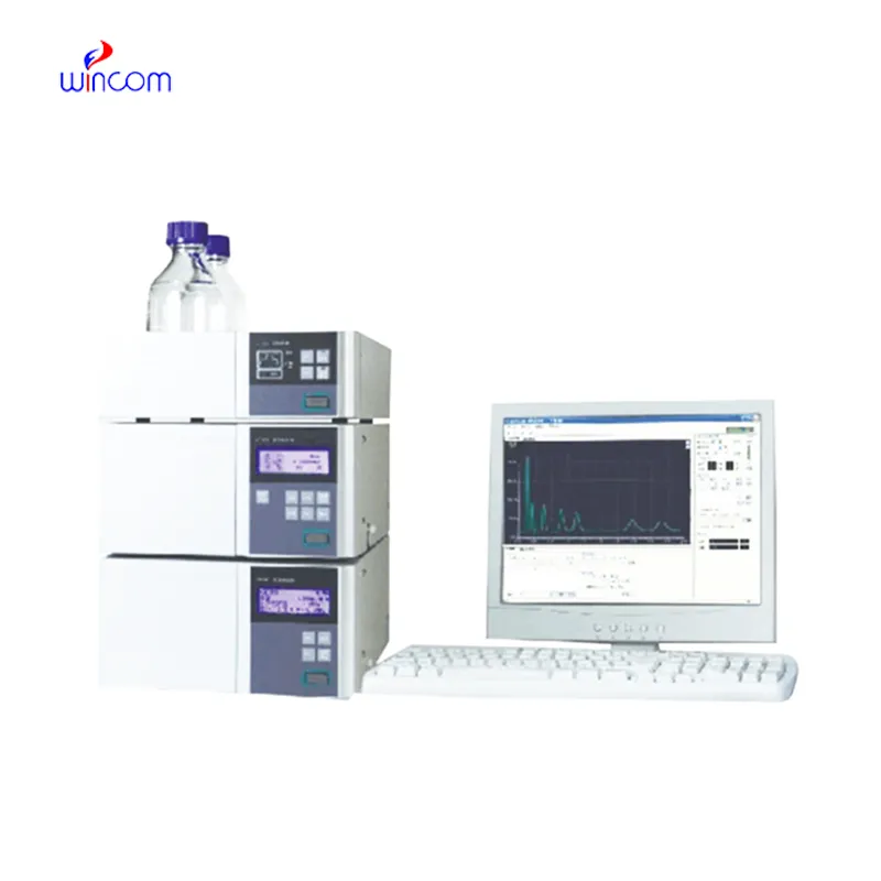
The hospital laboratory technicians employ stationary phase in hplc to quantify the quantity of proteins or peptides. This assists in the research of biomarkers, immunotherapy studies, and analysis of responses induced by novel therapies among patients. Its accuracy and sensitivity enable the obtaining of correct results, hence aiding superior research.
The instruments for stationary phase in hplc of the future will be equipped with separation methods in multiple dimensions and fully automated sample preparation. The detection of trace amounts of metabolites, drugs, and biomarkers will be so accurate that hospitals and clinical laboratories will be the first to reap the benefits. The applications of stationary phase in hplc in the future will greatly help in complex diagnostics, research studies, and laboratory efficiency.
The effectiveness of a laboratory is determined by the proper maintenance of stationary phase in hplc. If the pump seals are regularly cleaned, the flow rates are monitored, and the usage of incompatible solvents is avoided then damage to the laboratory equipment can be prevented. It is essential for the technicians to carefully examine the columns, detectors, and tubing and in case of any sign of wear to conduct the scheduled calibration. Keeping stationary phase in hplc in their best condition guarantees reproducibility, lowers the risk of equipment breakdown, and provides continuous performance for both hospital tests and experiments.
stationary phase in hplc is equipped with an in-depth examination of biomolecules like proteins, peptides, and nucleic acids. Reversed-phase, ion-exchange, and size-exclusion chromatography methods qualify scientists to get insight into the molecular properties with utmost accuracy. The application of stationary phase in hplc in metabolomics studies, enzyme kinetics, and protein characterization helps in high accuracy and reproducibility. The high sensitivity level helps to detect low-molecular-weight molecules in detail and get insight into biological samples at a high level. One of the prime reasons why scientists are interested in stationary phase in hplc is its ability to generate information that advances understanding at an advanced biochemistry level.
Q: What types of HPLC columns are available? A: Reversed-phase, normal-phase, ion-exchange, and size-exclusion columns are the main types of columns used according to the nature of the analytes. Q: Can multiple samples be analyzed simultaneously? A: Yes, in high-throughput systems, automated sample injection and sequential analysis are among the techniques to achieve this. Q: How does temperature affect HPLC performance? A: Temperature changes can cause variations in separation efficiency and retention times; however, the majority of labs make use of precise temperature control. Q: Can HPLC be integrated with data software? A: Sure, it can be linked with laboratory software for data collection, processing, and reporting. Q: What types of laboratories use HPLC? A: HPLC is employed by hospitals, pharmaceuticals, biochemistry research, and environmental testing labs.
This ultrasound scanner has truly improved our workflow. The image resolution and portability make it a great addition to our clinic.
We’ve been using this mri machine for several months, and the image clarity is excellent. It’s reliable and easy for our team to operate.
To protect the privacy of our buyers, only public service email domains like Gmail, Yahoo, and MSN will be displayed. Additionally, only a limited portion of the inquiry content will be shown.
Hello, I’m interested in your centrifuge models for laboratory use. Could you please send me more ...
We’re currently sourcing an ultrasound scanner for hospital use. Please send product specification...
E-mail: [email protected]
Tel: +86-731-84176622
+86-731-84136655
Address: Rm.1507,Xinsancheng Plaza. No.58, Renmin Road(E),Changsha,Hunan,China

 af
af
 es
es
 ar
ar
 tr
tr
 sw
sw
 pt
pt
 th
th
 ur
ur
 bn
bn
 ne
ne
 vi
vi
 km
km
 lo
lo
 de
de
 ru
ru
 fi
fi
 nl
nl
 fa
fa
 fr
fr
 ko
ko