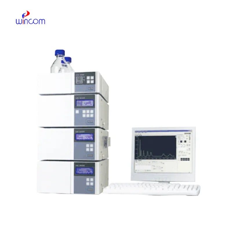
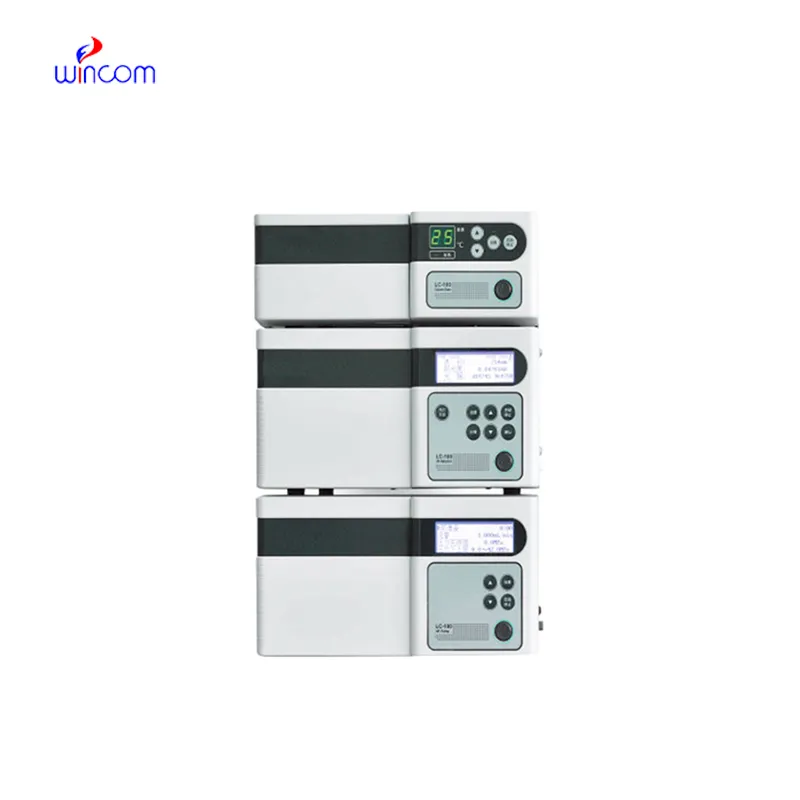
In the pharmaceutical lab, hplc instrument agilent is the key to the precise assessment of the active substances, impurities, and metabolites. The machine gives a high-resolution separation, which in turn supports the quality assurance and the regulation compliance. Lab workers put their trust on hplc instrument agilent for method validation, production consistency monitoring, and research trials. hplc instrument agilent brings together the delicate ability to detect plus the repeated nature of results to make the complex formulations proficiently analyzed, thus, it serves the routine lab testing and the advanced experimental work in hospitals, research centers, and clinical facilities both.
hplc instrument agilent is indispensable in the hospital lab for vitamin and nutrient analyses of patient samples. It identifies and determines the amounts of vitamins and minerals that are deficient or excessive in blood or serum. Health care providers depend on it to keep track of patients' nutrition, provide aids for treatment, and assess the impact of supplementation which, thus, boosts the quality of clinical care overall and makes it more beneficial.
hplc instrument agilent is expected to have an increasing role in personalized medicine, analyzing complicated biomarkers swiftly. In the future, their application in hospitals will be centered on integrating pharmacokinetics, metabolomics, and monitoring, helping medical practitioners have access to swift and comprehensive data. The workflow in laboratories is expected to be organized.
The effectiveness of a laboratory is determined by the proper maintenance of hplc instrument agilent. If the pump seals are regularly cleaned, the flow rates are monitored, and the usage of incompatible solvents is avoided then damage to the laboratory equipment can be prevented. It is essential for the technicians to carefully examine the columns, detectors, and tubing and in case of any sign of wear to conduct the scheduled calibration. Keeping hplc instrument agilent in their best condition guarantees reproducibility, lowers the risk of equipment breakdown, and provides continuous performance for both hospital tests and experiments.
hplc instrument agilent is of utmost importance in biochemistry laboratories of both universities and hospitals. It makes detailed study of proteins, peptides, and metabolites possible through the separation of intricate mixtures. The application of it includes but is not limited to enzymatic analysis, biomarker detection, and data obtained through metabolomics. The sensitivity and reproducibility of the device guarantee genuine molecular profiles. Lab technicians make use of hplc instrument agilent to conclude their experiments and provide evidence for scientific publications. Its accuracy and versatility give biochemistry labs the ability to perform cutting-edge research in molecular mechanisms, disease pathways, and therapy targets thus, it becomes an indispensable tool for both analytical and clinical lab investigations.
Q: What is the sample preparation for HPLC? A: For the most part, samples should be filtered, diluted, or subjected to solvent extraction in order to avoid column clogs and have the results be accurate Q: Is HPLC able to pick trace-level compounds? A: With the right detectors, it can pick up such substances in extremely small amounts with high sensitivity. Q: Is HPLC a method that can be applied to analysis of proteins? A: Yes, particularly if one employs size-exclusion and reversed-phase columns for protein, peptide, and biomolecule separation. Q: What is the process of calibrating HPLC? A: The process is done by taking standards of known concentrations that are the same as the one in the sample and using them to check the performance of the column and the accuracy of the detector. Q: Are particular solvents needed for HPLC? A: Yes, the solvents used need to be compatible with the type of the column and the detectors to prevent any damage or interference in the analysis process.
I’ve used several microscopes before, but this one stands out for its sturdy design and smooth magnification control.
We’ve used this centrifuge for several months now, and it has performed consistently well. The speed control and balance are excellent.
To protect the privacy of our buyers, only public service email domains like Gmail, Yahoo, and MSN will be displayed. Additionally, only a limited portion of the inquiry content will be shown.
We are planning to upgrade our imaging department and would like more information on your mri machin...
We’re interested in your delivery bed for our maternity department. Please send detailed specifica...
E-mail: [email protected]
Tel: +86-731-84176622
+86-731-84136655
Address: Rm.1507,Xinsancheng Plaza. No.58, Renmin Road(E),Changsha,Hunan,China

 af
af
 es
es
 ar
ar
 tr
tr
 sw
sw
 pt
pt
 th
th
 ur
ur
 bn
bn
 ne
ne
 vi
vi
 km
km
 lo
lo
 de
de
 ru
ru
 fi
fi
 nl
nl
 fa
fa
 fr
fr
 ko
ko